
mRetina AI
Telemedicine reimagined

Telemedicine reimagined
Back to Login
Back to Login
You have reached the maximum number of prompts allowed. Please contact support or upgrade your account for more access.
By using mRetinaAI, you agree to the following terms and conditions:
Ensure:
- A stable & fast internet connection
- An OCT image file in a supported format (e.g., JPEG, PNG, GIF, BMP)
- PDF, DOCx, and other document file types are not supported.
- The image should be clear, well-centered, and grayscale only.
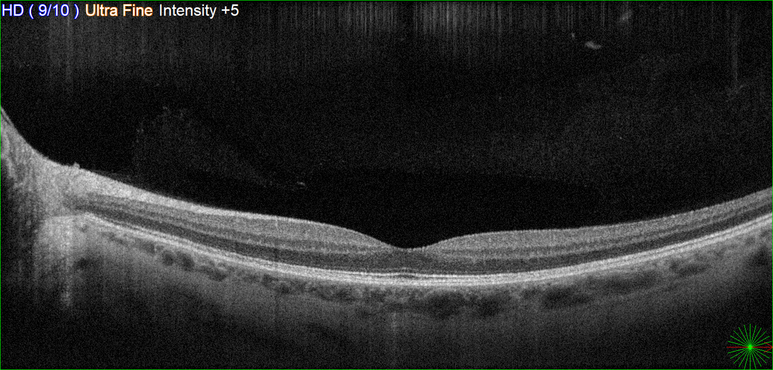
- Avoid images with excessive noise or artifacts.
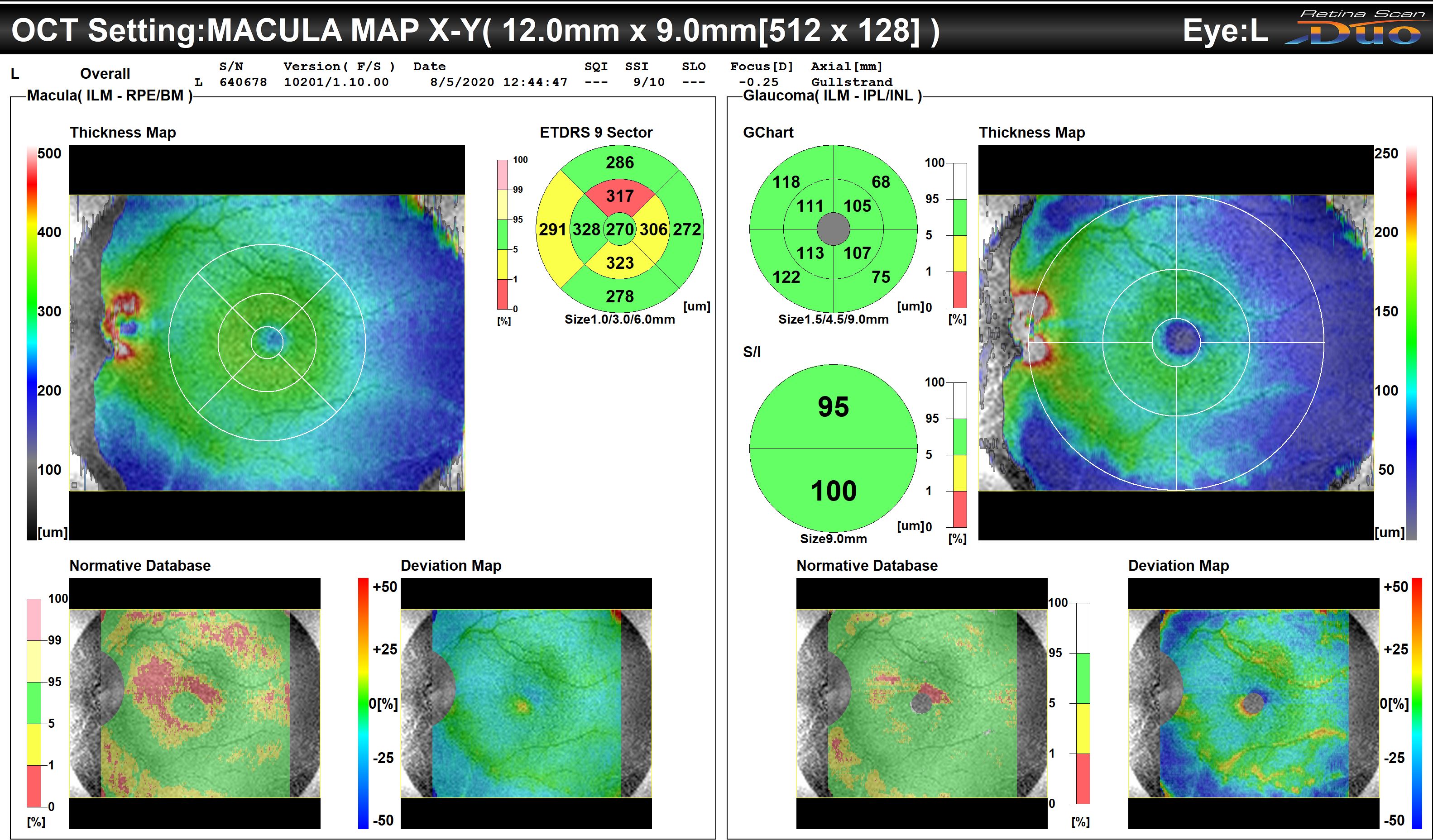
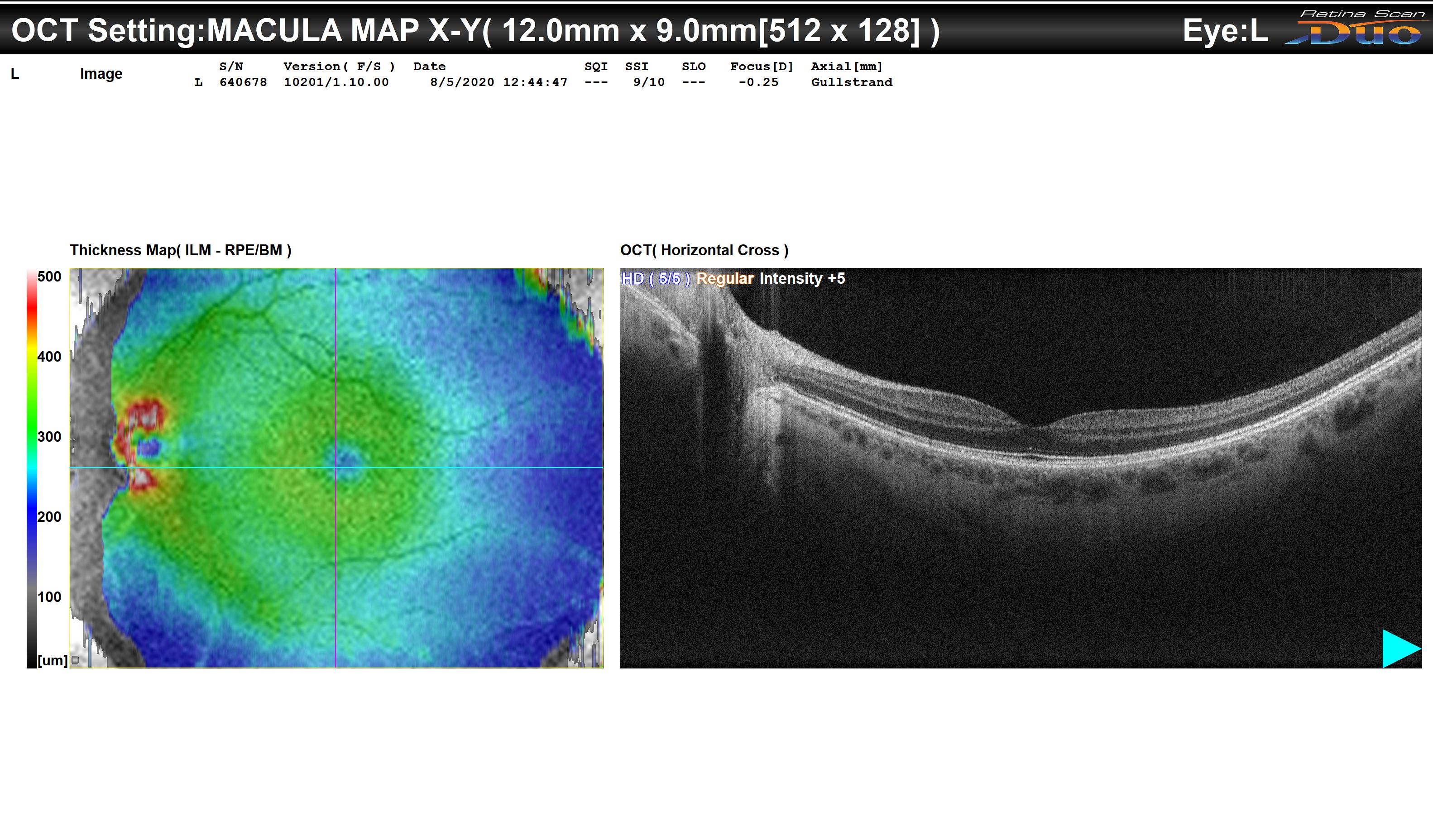
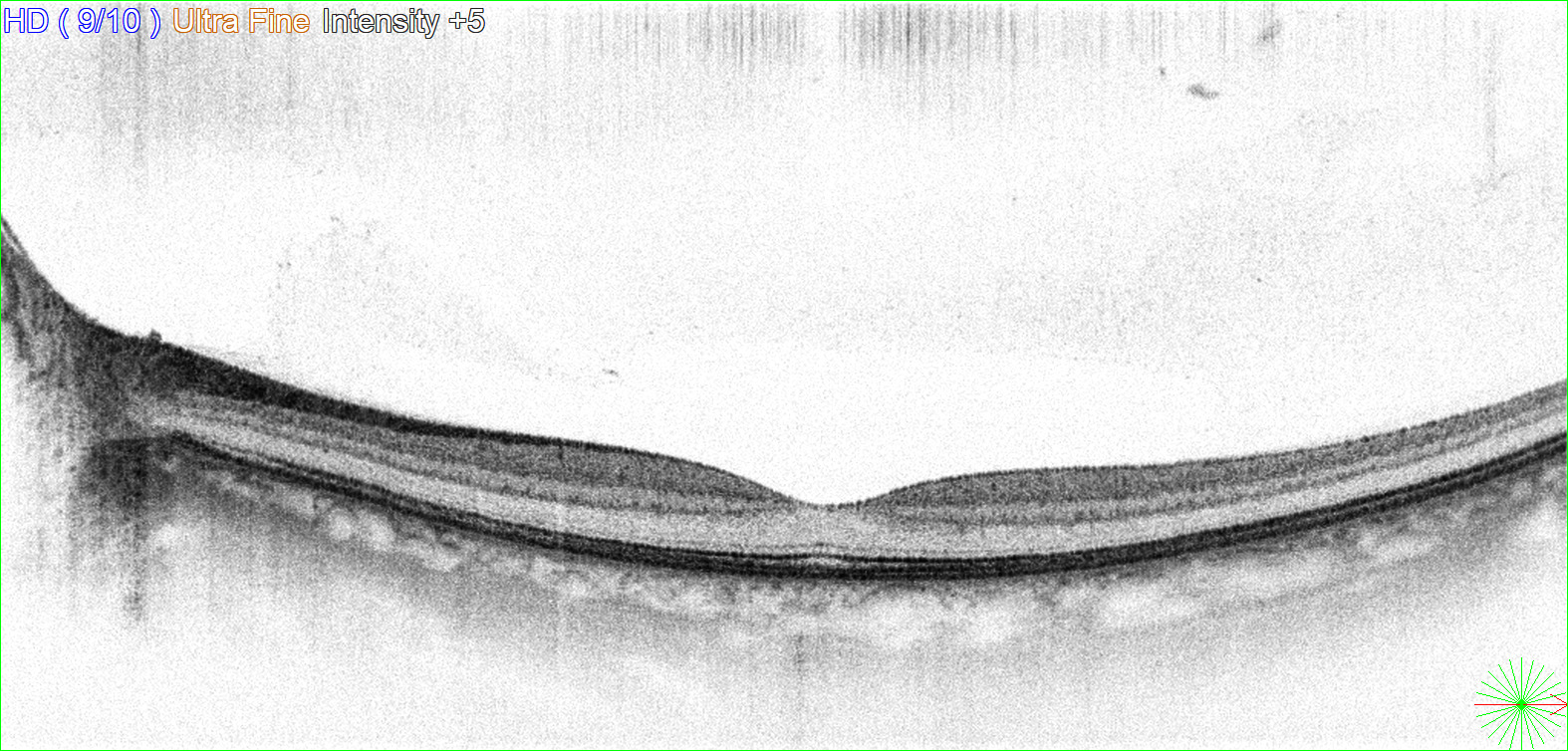
- Do not use images like Macular Map, Pseudo-color, Inverted grayscale, or those captured from a computer screen.
These are examples of images you should avoid:

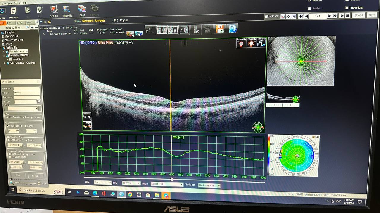
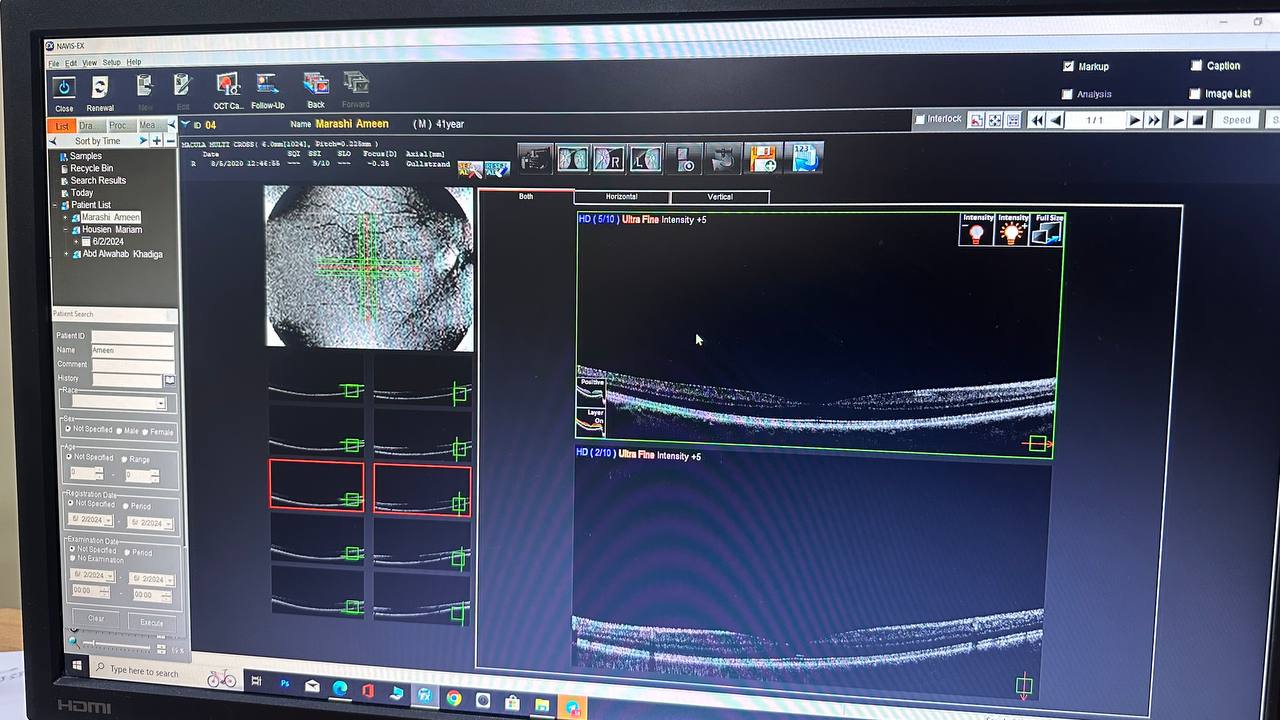
Using the Classifier:
Select the correct clinical entity:
- Upload your image by clicking on the designated area labeled "Drag and drop an OCT image file here, or click to select file."
- Once uploaded, the image will be displayed on the screen.
- Click "Predict" to analyze the image.
- The system will process the image and display the predicted condition.
- The image classifier is capable of predicting only specific pathologies based on the OCT images uploaded:
- The image classifier will not extract features from OCT images such as subretinal or intraretinal fluids.
- When diabetic retinopathy and macular edema are selected, the OCT image classifier can predict whether the uploaded image is Central DME, Non-central CSME, DME with Vitreomacular abnormalities, or Tractional Retinal Detachment.
- If Retinal Vascular Occlusive diseases are selected, the OCT image classifier can predict whether the uploaded image is a Long-Standing Retinal Vascular Occlusion, Macular Edema Related to Retinal Vein Occlusion, or Retinal Artery Occlusion.
- When AMD is selected for the OCT image classifier, it can predict whether the uploaded image is Non-neovascular AMD, Non-neovascular AMD with Geographic atrophy, or Active Neovascular AMD.
- If Pachychoroid is selected, the OCT image classifier can predict whether the uploaded image is CSCR, CSCR complicated with MNV, or Polypoidal Choroidal Vasculopathy.
- If Vitreo Macular Interface Abnormalities are selected, the OCT image classifier can predict whether the uploaded image is Lamellar Macular Hole, Vitreomacular traction, Full-thickness Macular Hole, or Epiretinal Membrane.
- When Hereditary Retinal Diseases are selected, the OCT image classifier can predict whether the uploaded image is Retinitis Pigmentosa, Retinitis Pigmentosa with Macular Edema, X-linked Juvenile Retinoschisis, or Vitelliform Lesion.
- If the predicted condition is not "Normal," click the "Suggest Recommendations" button to view related treatment options.
- Always verify the predictions before considering any recommendations.
- If you encounter an error, ensure the image file is in a supported format and try again.
- The classifier's accuracy is about 70% with correct images. Always ensure optimal image quality.
- If no prediction or recommendation button appears, try re-uploading the image or refreshing the page.